Program Progress:
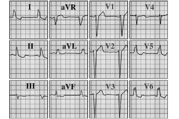
You are incorrect - our patient's electrocardiogram demonstrates left ventricular hypertrophy.
Click on the links to learn about this ECG:
Your choice: Complete left bundle branch block
The characteristic features demonstrated here include a wide QRS ≥ .12 seconds with mid QRS notching, absence of normal septal Q waves, and secondary ST-T wave changes. These abnormalities are seen in leads that best reflect the left ventricle, such as leads I, aVL, V5, and V6. The electrocardiographic changes seen in complete left bundle branch block often obscure other diagnostic changes, such as those seen with left ventricular hypertrophy and myocardial ischemia and/or infarction.
